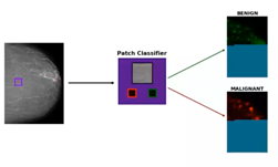
According to the National Institutes of Health (NIH), cervical cancer is third most common cancer among women and the second most frequent cause of cancer-related death, with 80% of cervical cancer cases occurring in developing nations with limited access to cervical cancer screening.
To help alleviate the problem, researchers from the NIH and intellectual Ventures Global Good Fund developed a deep learning-based proof-of-concept that can automatically evaluate cervical images for cancer.
“Human papillomavirus vaccination and cervical screening are lacking in most lower resource settings, where approximately 80% of more than 500 000 cancer cases occur annually,” the researchers stated in their paper. “The objective of this study was to develop a “deep learning”-based visual evaluation algorithm that automatically recognizes cervical precancer/cancer.”
Using NVIDIA Tesla V100 GPUs on a DGX-1, with the cuDNN-accelerated TensorFlow and Keras deep learning frameworks, the team trained a convolutional neural network on approximately 30,000 digitized cervical images from screening, taken with a fixed-focus camera (“cervicography”) from more than 9000 participants. With the help of the Faster R-CNN algorithm, as well as extensive image augmentation, the system learned to detect the cervix and report the probability of cancer.
“This longitudinal cohort study of HPV and cervical cancer was conducted in a random sample of a moderate-risk, poorly screened Costa Rican province,” the researchers said. “Participants aged 18–94 years were screened at baseline and periodically for up to 7 years using multiple methods at intervals determined by their screening results and resultant estimated risk of precancer.”
According to the researchers, Each visit included multiple kinds of tests: cytology, HPV testing, and cervicography.
“The proof-of-principle strongly supports further evaluation of automated visual evaluation,” the researchers said. “Rather than resurrecting obsolete film camera technology to achieve the observed results, we are currently working to transfer automated visual evaluation to images from contemporary phone cameras and other digital image capture devices to create an accurate and affordable point-of-care screening method that would support the recently announced World Health Organization initiative to accelerate cervical cancer control.”
Even though the work is still in the research phase, the automated visual evaluation system identified cumulative precancer/cancer cases with greater accuracy than original cervigram interpretation or conventional cytology. And despite the input to this CNN being images from Cervicography, a procedure that is not used in current practice, the researchers hope that in the future their work can be used for automated visual evaluation of cervical images from contemporary digital cameras, to help patients with effective point-of-care cervical screening..
The research was recently published in the Journal of the National Cancer Institute.
Read more>
NIH Develops an AI to Help With Cervical Cancer Screening
Jan 24, 2019
Discuss (0)

Related resources
- GTC session: How Artificial Intelligence is Powering the Future of Biomedicine
- GTC session: Creating AI-Powered Hardware Solutions for Medical Imaging Applications
- GTC session: Generative AI Theater: 15 Minutes to Change the World: Generative AI in Healthcare
- NGC Containers: VISTA-3D NIM
- NGC Containers: MATLAB
- NGC Containers: BodyMarker/PhenoBody