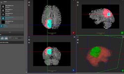
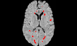
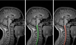
A team of researchers from some of the top medical institutions in the world, developed a fully automatic deep learning-based system to detect multiple sclerosis (MS) lesions in the spinal cord and intramedullary from conventional MRI data.
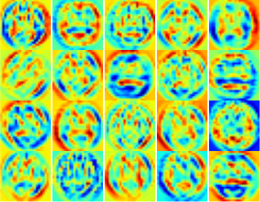
MS is a chronic immune disease of the central nervous system which appears in areas of the brain and spinal cord with the occurrence of focal regions of inflammatory demyelination. To diagnose the disease and track its progress, clinicians and radiologists use MRIs to detect the lesions, a process that is time-consuming and suffers from variability.
The study, conducted by the University of Montreal, San Francisco General Hospital, Massachusetts General Hospital, the National Institute of Health (NIH), Stanford University, Vanderbilt University, Harvard Medical School, and many other institutions, has the potential to radically alter how MS is detected and tracked.
Using NVIDIA Tesla P100 GPUs, and the cuDNN-accelerated Keras and TensorFlow deep learning frameworks, the researchers trained a convolutional neural network on MRI images of 1,042 patients to automatically detect lesions in both the spinal cord and intramedullary.
“Segmentation of the spinal cord and lesions from MRI data provides measures of atrophy and lesion burden, which are key criteria for the diagnosis, prognosis and longitudinal monitoring in MS. Achieving robust and reliable segmentation across multi-center spinal cord data is challenging because of the large variability related to acquisition parameters and image artifacts,” the researchers wrote in their research paper. “There is a need for robust and automatic segmentation tools for the spinal cord and the MS lesions within.”
The spinal cord segmentation results outperformed a state-of-the-art method and the other lesion segmentation performed within the range of comparable to humans.
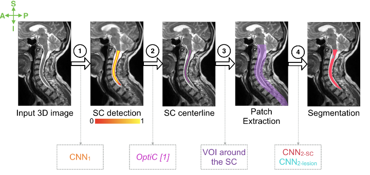
“When compared to a state-of-the-art spinal cord segmentation method (PropSeg), our CNN-based approach showed a median Dice of 95% vs. 88% for PropSeg. Regarding lesion segmentation, our framework, when compared with manual segmentation of MS patients, provided a lesion-wise detection sensitivity of 83%, a precision of 77%, a relative volume difference of 15%, and a Dice of 60%” the researchers said.
The MS lesion segmentation method was based on a cascade of two convolutional neural networks, which the researchers have publicly released.
The research was published on ArXiv earlier this month and was submitted to the NeuroImage Journal.
Read more >
AI Helps Doctors Detect MS In the Spinal Cord
May 21, 2018
Discuss (0)
Related resources
- DLI course: Image Classification with TensorFlow: Radiomics?€?1p19q Chromosome Status Classification
- DLI course: Data Augmentation and Segmentation with Generative Networks for Medical Imaging
- GTC session: Creating AI-Powered Hardware Solutions for Medical Imaging Applications
- GTC session: Optimizing Your AI Strategy to Develop and Deploy Novel Deep Learning Models in the Cloud for Medical Image Analysis
- GTC session: How Artificial Intelligence is Powering the Future of Biomedicine
- SDK: MONAI Deploy App SDK